

Overview
Documents
Training
Advanced Bone Density Assessment for Confident Clinical Decisions
Dual-energy X-ray Absorptiometry (DXA) is the gold-standard technology for measuring bone mineral density and assessing fracture risk. Our DXA system delivers fast, precise, and low-dose scans, helping clinicians detect osteoporosis early, monitor treatment over time, and support proactive bone health management for every patient.
Powerful images. Clear answers.
Fast Exams
Bone density scans are completed in about 10–30 seconds1, and body composition assessments as little as 2 minutes2.
Patient-focused design
Supports patients weighting up to 500 pounds and offers 24-inch C-arm clearance to help accommodate a wide range of body types and mobility needs.
Long term precision
Horizon delivers pixel-by-pixel calibration through bone and tissue for greater long-term measurements stability12.
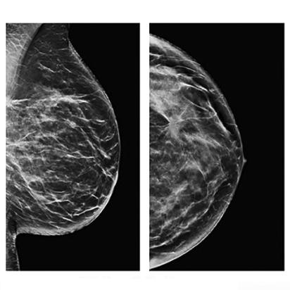
Exceptional Image Quality
OnePass™ single sweep scanning designed to eliminate beam overlap errors and image distortion.
Comprehensive Clinical Applications with DXA
Osteoporosis assessment is the process of evaluating bone health and bone mineral density to understand an individual’s risk of weakened bones and fractures.

Osteoporosis is one of the most under diagnosed and under treated threats to lifelong mobility1:
- 1 in 2 women over age 50 will break a bone due to osteoporosis.2
It affects 8 million women and 2 million men in the U.S.3
DXA is the gold standard for osteoporosis assessment. Our Hologic Horizon® DXA delivers:
- A quick scan of only 10-30 seconds4
- High-precision bone density measurements to help detect osteoporosis before fractures occur5
- Advanced fracture risk insights to support early, confident treatment decisions5
For qualified individuals, DXA exams are typically covered by insurance every 2 years.6
Patients can visit findmydxa.com to learn more about how osteoporosis can affect their daily life, understand what to expect from a bone density (DXA) exam, and easily find a nearby facility.
Body composition assessment is the process of evaluating the proportions of fat, muscle, bone, and other tissues that make up a person’s body.

In just 2 minutes1, obtain up to 220 precise, regional measurements2 of fat, lean mass, and bone to help:
- Evaluate the success of fitness and sports interventions3
- Optimize training and performance programs
- Identify muscle imbalances that may increase injury risk
- Support confident, data-driven return-to-play decisions
Backed by extensive clinical evidence, this DXA-based solution incorporates reference data from more than 20,000 individuals in the CDC’s NHANES (National Health and Nutrition Examination Survey) study, delivering reproducible, research-grade insights you can trust.4
For deeper performance analytics, segmental reporting, and sports-specific use cases, visit our dedicated Body Composition website at dxaperformance.com.
Breast Cancer and Bone Density
Breast cancer treatment can significantly impact bone health—patients may lose 6–7% of bone density during therapy13. Aromatase inhibitors, a common endocrine therapy for postmenopausal patients with HR+, early-stage breast cancer, can further accelerate bone loss and increase fracture risk.
The Breast Cancer Index® test helps determine whether a patient is likely to benefit from extending endocrine therapy beyond 5 years. When extended therapy is unlikely to help, she may be able to avoid additional exposure to bone toxicity and its impact on long-term bone health.